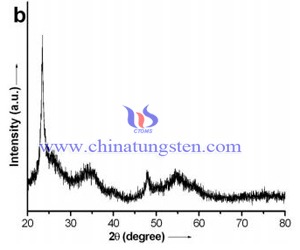
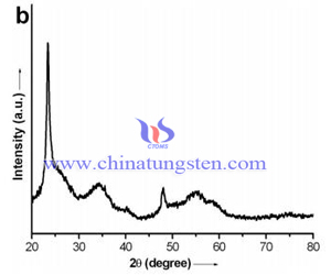
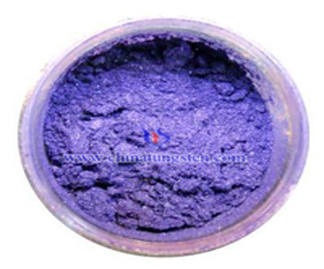
Violet Tungsten Oxide XRD Photo
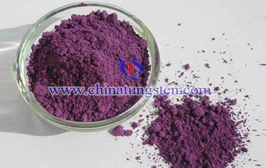
Violet tungsten oxide XRD photo is using nature electromagnetic wave of X-ray to carry out diffraction and then getting the related image. The picture following is the typical violet tungsten oxide XRD photo, wherein the built-in figure is the molecular structure model image.

X-ray diffractometer is a typical diffractometer consists of a source of radiation, a monochromator to choose the wavelength, slits to adjust the shape of the beam, a sample and a detector. In a more complicated apparatus, a goniometer can also be used for fine adjustment of the sample and the detector positions.
There are several types of of X-ray diffractometer, depending of the research field (material sciences, powder diffraction, life sciences, structural biology, etc.) and the experimental environment, if it is a laboratory with its home X-ray source or a Synchrotron. In laboratory, diffractometers are usually a "all in one" equipment, including the diffractometer, the video microscope and the X-ray source. Plenty of companies manufacture "all in one" equipment for X-ray home laboratory, such has Bruker, Rigaku, PANalytical, Thermo Fisher Scientific and many others.
